Файл:SARS-CoV-2 PHIL23591.png

Содержимое страницы недоступно на других языках.
Материал из Викиновостей, свободного источника новостей

Исходный файл(1461 × 1461 пкс, размер файла: 2,46 МБ, MIME-тип: image/png)

Краткое описание

Описание
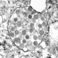
English: Thin section electron microscopic image of SARS-CoV-2, the causative agent of COVID-19. Spherical virus particles contain black dots, which are cross-sections through the viral nucleocapsid. In the cytoplasm of the infected cell, clusters of particles are found within the membrane-bound cisternae of the rough endoplasmic reticulum/Golgi area.
Дата
Источник

This media comes from the Centers for Disease Control and Prevention's Public Health Image Library (PHIL), with identification number #23591.

Note: Not all PHIL images are public domain; be sure to check copyright status and credit authors and content providers.


العربية | Deutsch | English | македонски | slovenščina | +/−

Автор Cynthia S Goldsmith and Azaibi Tamin; CDC

Лицензирование

Public domain
Это изображение было создано или получено агентством Центры по контролю и профилактике заболеваний, которое является подразделением Министерства здравоохранения и социальных служб США. Это было сделано при выполнении служебных обязанностей государственным служащим. Поскольку изображение создано Федеральным правительством США, оно находится в общественном достоянии (public domain).

eesti  Deutsch  čeština  español  português  English  français  Nederlands  polski  slovenščina  suomi  македонски  українська  日本語  中文(简体)‎  中文(繁體)‎  العربية  +/−

Краткие подписи

Добавьте однострочное описание того, что собой представляет этот файл
virus

image/png

История файла

Нажмите на дату/время, чтобы увидеть версию файла от того времени.

Дата/времяМиниатюраРазмерыУчастникПримечание
текущий17:24, 1 июня 2020Миниатюра для версии от 17:24, 1 июня 20201461 × 1461 (2,46 МБ)-sasha-== {{int:filedesc}} == {{Information |Description={{en|Thin section electron microscopic image of SARS-CoV-2, the causative agent of COVID-19. Spherical virus particles contain black dots, which are cross-sections through the viral nucleocapsid. In the cytoplasm of the infected cell, clusters of particles are found within the membrane-bound cisternae of the rough endoplasmic reticulum/Golgi area.}} |Source={{CDC-PHIL|23591}} |Date=2020 |Author=Cynthia S Goldsmith and Azaibi Tamin; CDC |Permis...

Следующая страница использует этот файл:

Метаданные